Author: Michael

  • WSL-1800CytoWatcher

    WSL-1800 CytoWatcher is compact digital microscope.

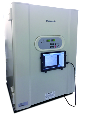
    No stress to take space in laboratory and moisture resistant for setting in COincubator to detect time lapse live cell.
    WSL-1800 CytoWatcher
    Purpose and Application

    To detect bright field images for culture cell, stained cell/tissue, time-lapse live cell.

    Usable like a microscope.

    Product Summary

    WSL-1800 CytoWatcher is applied a 5M pixels with color digital camera.

    The camera is compatibility to detecting a cell, tissue etc.

    CytoWatcher is simple to install only connect USB cable with PC and control software is able to detect an image, save an image, create movie file etc.

    CytoWatcher is moisture resistant and usable in incubator, clean bench in order to detecting a time-lapse live cell.

    Features

    Time-lapse live cell imaging with incubating

    Set CytoWatcher at incubator and detected in time-lapse live cell

    (Auto contrast:ON, created movie file)
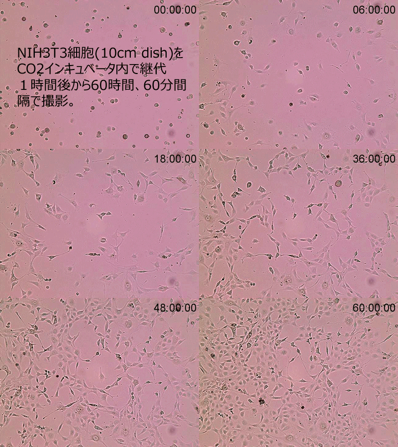
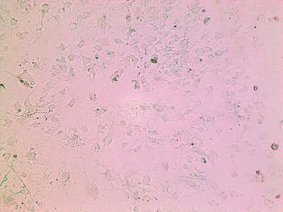
    NIH3T3_GIF動画

      Cultured  NIH3T3 cells in 10cm dish

      Time-lapse: 60 min intervals for 60 hours in COincubator

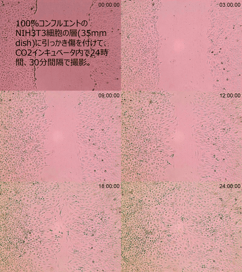
    Healing-Assay

      Wound healing assay with NIH3T3 cells in 35mm dish

      Time-lapse: 30 min intervals for 24 hours in COincubator.

    5M Pixels color digital camera

    CytoWatcher is applied 4x object lens with 5M pixels color digital camera.

    Field of view 1.425mm×1.070mm
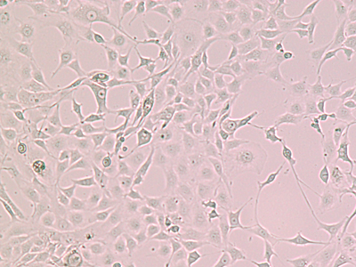
    1_4

      1/4 Scale down

    1_2

      1/2 Scale down

    1_1

      Display 100%(Monitor resolusion 1:1)

    Contrast adjustment

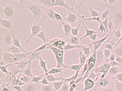
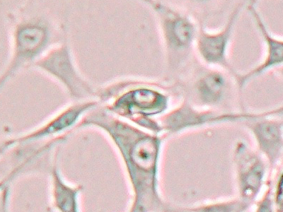
    Transparent cell case, difficult to see it because of low contrast.

    Cytowatcher control software can catch easy to see it by auto-contrast adjusting function.
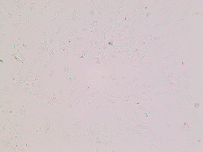
    コントラスト調整前

      Before contrast adjustment

    コントラスト調整後

      Auto-contrast adjusted image

    Imaging of Stained Cells
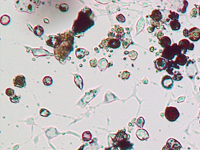
    色素染色画像1_2

      Adipose differentiated cells(mouse Swiss 3T3 L-1 derivered)

      Oil Red O stain
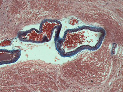
    切片画像

      Section preparation of basal ganglia(mammal)

      Masson trichrome stain

    Specifications
    Camera5M Pixels Color CMOS
    Resolusion2592×1944 pixels 
    Object Lens 4x
    FocusManual adjustment
    Field of view1.425mm×1.070mm
    Light sourceWhite LED
    Moisture ResistantUsable humidity 95%RH(Usable in COincubator)
    Control SoftwareImageSaver6 for Windows
    Detection method: Live/Still image/Time lapsed
    Save file: 8bit TIFF/JPEG/BMP(Color/Monochrome)
    PC connection USB 3.0 (Monitor resolution is needed more than 1024×768)
    Cable length 0.8m(80cm)※
    OSWindows10/8.1/7 64bit/32bit
    Size130mm(W)×180mm(D)×190mm(H)
    Weight2.5kg
    PowerUSB(bus power)DC5V

    ※Extension USB cable is option 「No.3601809 USB3.0 extension cable」

    Brochure

    Download Catalog PDF

    WSL-1800 CytoWatcher brochure

    Control software

    Installation a control software「ImageSaver6」 to Windows PC(connection with USB3.0 cable)

    Detect an image for live, still image, time-lapse etc.
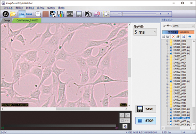
    CytoWatcherソフト画面(2)Live細胞4x拡大

    Usable like a small microscope

    CytoWatcher is usable a petri dish, slide like microscope.  
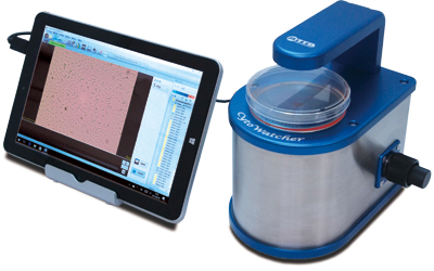
    WSL-1800_2017_左向きPCシャーレ付①

    Usable in Clean bench

    Usable in clean bench with option USB3.0 extension cable
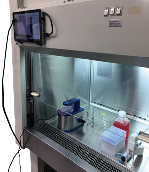
    WSL-1800_2017_クリーンベンチ内①のコピー

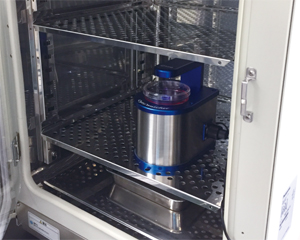
    Detection a time-lapse in incubator

    CytoWatcher is moisture resistance device. 
    WSL-1800_2017_インキュベータ内④
    WSL-1800_2017_インキュベータ内⑦
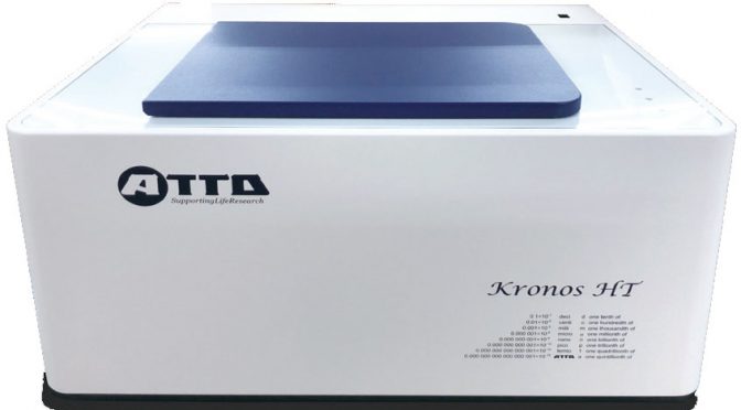
  • WSL-1565 Kronos HT

    WSL-1565 Kronos HT

    NEW WSL-1565 Kronos HT Brochure 2019

    Download (PDF, 84.55MB)

    Kronos HT main unit, Control software (for Windows PC), CO2gas mixer, Humidifying unit, 24 well plate adapter

    • High throughput live cell realtime reporter assay
    • Measuring with temperature and CO2 controlled incubation chamber
    • 24 well plate x 2 or 96 well plate x 2 (option)
    • Data display with real-time
    • Detectors scan each well, so culture plates don’t move.
    • Lower noise by PMT cooling
    • Dual-color luciferase assay by optical filter auto-changing

    Detector: Photomultiplier tube (10oC cooling) x 2 units, X-Y actuating
    Measurement: Photon counting method
    Measurement time: Integral time: 1 – 60 s/well, Measurement term: 1 h – 30 days
    Plate format: Clear bottom 24 well plate x 2 (Standard)
    Clear bottom 96 well plate x 2 (Option), 35 mm dish x 12 (Option)
    Throughput:
    48 samples (24 well plate x 2)
    192 samples (96 well plate x 2, option)
    12 samples (35 mm dish x 12, option)
    Incubation temperature control: Room temperature + 5oC to 45oC at room temperature 20 – 28oC
    CO2 gas control: Control to 5% by CO2 gas mixer
    Humidification:
    Setting of water reservoir in incubator and humidified 5% CO2 gas flowing intermittently by
    humidifying unit
    Maintaining >90%RH
    Multi-color assay: Measurement with automatically selected optical filter up to 2 colors
    PC requirements OS: Windows 10 / 8.1 / 7, 64 / 32 bit, Memory: 4 GB, HDD: more than 20 GB free space
    Interface: USB2.0 x 1 port
    Dimensions, Weight:
    Main unit: 650 (W) x 520 (D) x 340 (H) mm, 40 kg
    CO2 gas mixer: 160 (W) x 300 (D) x 200 (H) mm, 4.8 kg
    Humidifying unit: 170 (W) x 140 (D) x 115 (H) mm, 1.6 kg
    Power:
    Main unit: AC 100 – 240 V, 50 / 60 Hz, 400 W (max)
    CO2 gas mixer: AC 100 – 240 V, 50 / 60 Hz, 100 W (max)
    Humidifying unit: AC 100 – 240 V, 50 / 60 Hz, 24W (max)

    Model 3510146: 24 well plate adapter
    Model 3510147: 96 well plate adapter
    Model 3510148: 35 mm dish adapter

    Application data

    Circadian rhythm of clock gene expression

    Transcriptional activity of clock gene, mPer2
    mPer2 promoter – Eluc (PEST) expressed fibroblast stable
    cell line in 24 well clear bottom plate
    Measurement time: 5s / well, Interval time: 10min

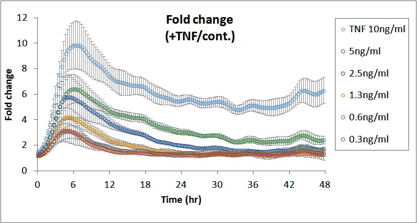
    Drug response of transcription factor

    Dual-color real-time luciferase assay of TNF-a induced NF-kB transcriptional activity

    NF-kB response elemnt – TK promoter – SLG (green color emited luciferase) and TK promoter – SLR (red color emitted luciferase)
    expressed fibroblast cell line in 96 well clear bottom plate
    Measurement time: 5s x 2 color / well, Interval time: 30min
    (A), (B) Transcriptional activity of NF-kB and TK promoter for 48h
    (C) Normalized by activity of control (TK)
    (D) Ratio to NF-kB activity without TNF-a

  • WSE-5200 Printgraph 2M

    WSE-5200 Printgraph 2M is a GDS with a high resolution monochrome CCD camera that could be operated by a touch control panel, or using Wi-Fi with a tablet. For a detection, it uses UV light source, white LED light source, Cyan LED and Red LED. Cyan LED is available to detect a fluorescence in both Blue color and Green color. Detectable EtBr which is difficult to detect in Blue LED. The UV light source could be turned off if preferred. The image is saved in main unit memory or USB memory, and if desired, it could be connected to UP-X898MD that printout instantly.

    Download (PDF, 3.48MB)