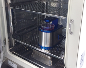
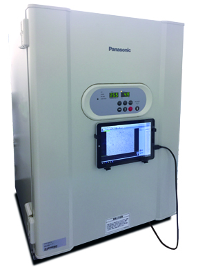
WSL-1800 CytoWatcher is compact digital microscope.
No stress to take space in laboratory and moisture resistant for setting in CO2 incubator to detect time lapse live cell.
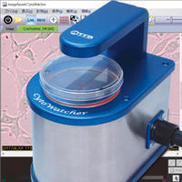
Purpose and Application
To detect bright field images for culture cell, stained cell/tissue, time-lapse live cell.
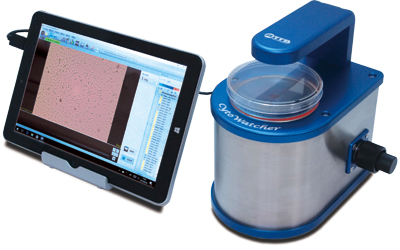
Usable like a microscope.
Product Summary
WSL-1800 CytoWatcher is applied a 5M pixels with color digital camera.
The camera is compatibility to detecting a cell, tissue etc.
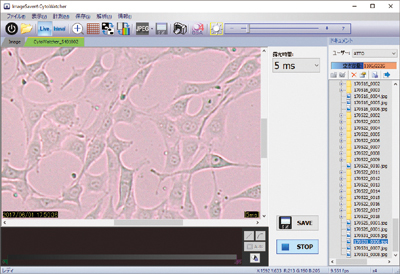
CytoWatcher is simple to install only connect USB cable with PC and control software is able to detect an image, save an image, create movie file etc.
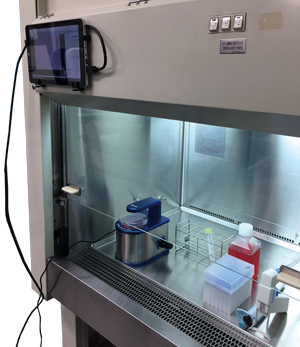
CytoWatcher is moisture resistant and usable in incubator, clean bench in order to detecting a time-lapse live cell.
Features
Time-lapse live cell imaging with incubating
Set CytoWatcher at incubator and detected in time-lapse live cell
(Auto contrast:ON, created movie file)
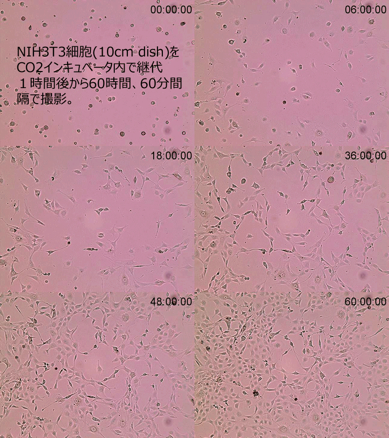
Cultured NIH3T3 cells in 10cm dish
Time-lapse: 60 min intervals for 60 hours in CO2 incubator
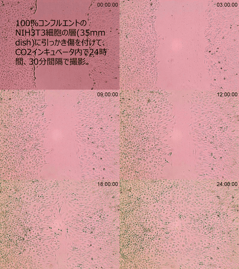
Wound healing assay with NIH3T3 cells in 35mm dish
Time-lapse: 30 min intervals for 24 hours in CO2 incubator.
5M Pixels color digital camera
CytoWatcher is applied 4x object lens with 5M pixels color digital camera.
Field of view 1.425mm×1.070mm
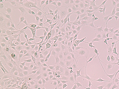
1/4 Scale down
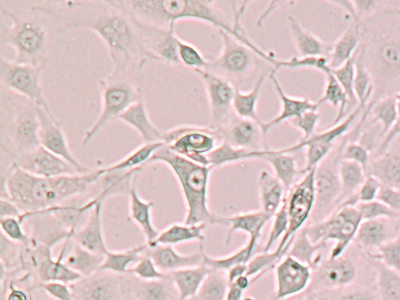
1/2 Scale down
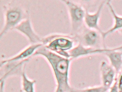
Display 100%(Monitor resolusion 1:1)
Contrast adjustment
Transparent cell case, difficult to see it because of low contrast.
Cytowatcher control software can catch easy to see it by auto-contrast adjusting function.
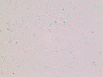
Before contrast adjustment
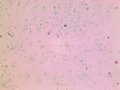
Auto-contrast adjusted image
Imaging of Stained Cells
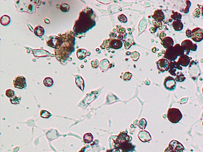
Adipose differentiated cells(mouse Swiss 3T3 L-1 derivered)
Oil Red O stain
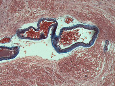
Section preparation of basal ganglia(mammal)
Masson trichrome stain
Specifications
| Camera | 5M Pixels Color CMOS |
| Resolusion | 2592×1944 pixels |
| Object Lens | 4x |
| Focus | Manual adjustment |
| Field of view | 1.425mm×1.070mm |
| Light source | White LED |
| Moisture Resistant | Usable humidity 95%RH(Usable in CO2 incubator) |
| Control Software | ImageSaver6 for Windows Detection method: Live/Still image/Time lapsed Save file: 8bit TIFF/JPEG/BMP(Color/Monochrome) |
| PC connection | USB 3.0 (Monitor resolution is needed more than 1024×768) Cable length 0.8m(80cm)※ |
| OS | Windows10/8.1/7 64bit/32bit |
| Size | 130mm(W)×180mm(D)×190mm(H) |
| Weight | 2.5kg |
| Power | USB(bus power)DC5V |
※Extension USB cable is option 「No.3601809 USB3.0 extension cable」
Brochure

WSL-1800 CytoWatcher brochure
Control software
Installation a control software「ImageSaver6」 to Windows PC(connection with USB3.0 cable)
Detect an image for live, still image, time-lapse etc.
Usable like a small microscope
CytoWatcher is usable a petri dish, slide like microscope.
Usable in Clean bench
Usable in clean bench with option USB3.0 extension cable
Detection a time-lapse in incubator
CytoWatcher is moisture resistance device.